43 eye diagram with labels and functions
The Human Eye - Diagram, Parts, Working, Function and Work of The Lens The cornea, iris, pupil, and lens make up the front of the eye, which focuses the image onto the retina. The light-sensitive membrane that covers the back of the eye is known as the retina. This membrane is made up of millions of nerve cells that clump together behind the eye to form the optic nerve, a huge nerve. The Human Eye › help › commGenerate eye diagram - MATLAB eyediagram - MathWorks eyediagram(x,n) generates an eye diagram for signal x, plotting n samples in each trace. The labels on the horizontal axis of the diagram range between –1/2 and 1/2. The function assumes that the first value of the signal and every nth value thereafter, occur at integer times.
Excel Gauge Chart Template - Free Download - How to Create Move the labels to the appropriate places above the gauge chart. Change the chart title. Bonus Step for the Tenacious: Add a text box with your actual data value. Here is a quick and dirty tip on making the speedometer chart more informative as well as pleasing to the eye. Let’s add a text box that will display the actual value of the pointer.

Eye diagram with labels and functions
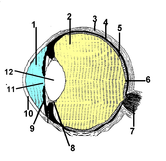
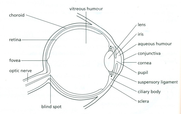
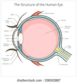
Eye Diagram With Labels and detailed description - BYJUS A brief description of the eye along with a well-labelled diagram is given below for reference. Well-Labelled Diagram of Eye The anterior chamber of the eye is the space between the cornea and the iris and is filled with a lubricating fluid, aqueous humour. The vascular layer of the eye, known as the choroid contains the connective tissue. Anatomy of the eye: Quizzes and diagrams | Kenhub Take a look at the diagram of the eyeball above. Here you can see all of the main structures in this area. Spend some time reviewing the name and location of each one, then try to label the eye yourself - without peeking! - using the eye diagram (blank) below. Unlabeled diagram of the eye. Click below to download our free unlabeled diagram of ... Eye Anatomy | Definition, Structure & Functions - iBiologia Diagram of Human Eye with Labelling. Eye Anatomy Complete Physiology of Eye is described below in the given paragraph: The eye is rather like a living Camera. Each eye is a liquid-filled ball 2.5 cm in diameter. At the front of the eye is a clear, round window called the cornea. Behind the cornea is a "lens.
Eye diagram with labels and functions. diagram of eye with labelling Eye Diagram With Labels And Functions - Aflam-Neeeak aflam-neeeak.blogspot.com Earthworm Presentation earthworm labeled dissection external anatomy labels presentation lumbricus label section cross slideshare sp fig physiology Labeled Eye Diagram ks2 links markcritz Human Eye Anatomy Quiz Labelled Diagram of Human Eye, Explanation and Function - VEDANTU The basic functions of Rods and Cones are conscious light perception, color differentiation and depth perception. The human eye is capable of distinguishing between about 10 million colors, and it can also detect a single photo. The human eye is a part of the sensory nervous system. Labeled Diagram of Human Eye byjus.com › biology › human-heartHuman Heart - Anatomy, Functions and Facts about Heart - BYJUS Practice your understanding of the heart structure. Drag and drop the correct labels to the boxes with the matching, highlighted structures. Instructions to use: Hover the mouse over one of the empty boxes. One part in the image gets highlighted. Identify the highlighted part and drag and drop the correct label into the same box. Eye anatomy and function - AboutKidsHealth For people with normally functioning eyes, the following sequence takes place: Light reflects off the object we are looking at. Light rays enter the eye through the cornea at the front of the eye. The light passes through a watery fluid (aqueous humor), and enters the pupil to reach the lens.
Human Eye Diagram, How The Eye Work -15 Amazing Facts of Eye First, light rays enter the eye through the cornea, the clear front "window" of the eye. The dome shaped cornea bends light to help the eye focus. From the cornea, the light passes through an opening called the pupil. The amount of light passing through is controlled by the iris, or the colored part of your eye. Liver Diagram with Detailed Illustrations and Clear Labels - BYJUS Liver – Anatomy, Functions, And Liver Diseases; Also Read: 6 Facts Everyone Should Know About The Liver; Fatty Liver Symptoms – Explore The Signs, Indications And Causes; 12 Alarming Symptoms of Liver Problems You Shouldn’t Ignore; A Brief Account Of Hepatic Portal System And Its Significance; Human Body – Anatomy and Physiology of ... PDF Parts of the Eye - National Institutes of Health To understand eye problems, it helps to know the different parts that make up the eye and the functions of these parts. Here are descriptions of some of the main parts of the eye: ... Handout illustrating parts of the eye Keywords: parts of the eye, eye diagram, vitreous gel, iris, cornea, pupil, lens, optic nerve, macula, retina ... en.wikipedia.org › wiki › Human_eyeHuman eye - Wikipedia Each eye has seven extraocular muscles located in its orbit. Six of these muscles control the eye movements, the seventh controls the movement of the upper eyelid.The six muscles are four recti muscles – the lateral rectus, the medial rectus, the inferior rectus, and the superior rectus, and two oblique muscles the inferior oblique, and the superior oblique.
Eye Diagram - an overview | ScienceDirect Topics An eye diagram provides a simple and useful tool to visualize intersymbol interference between data bits. Figure 24a shows a perfect eye diagram. A square bit stream (i.e., series of symbol '1's and '0's) is sliced into sub-bit stream with predetermined eye intervals (i.e., several bit periods), and displayed through bit analyzing equipment (e.g., digital channel analyzer), overlapping ... Human eye - Wikipedia The human eye is a sensory organ, part of the sensory nervous system, that reacts to visible light and allows us to use visual information for various purposes including seeing things, keeping our balance, and maintaining circadian rhythm.. The eye can be considered as a living optical device.It is approximately spherical in shape, with its outer layers, such as the outermost, white … Labelling the eye — Science Learning Hub The human eye contains structures that allow it to perceive light, movement and colour differences. In this activity, students use online or paper resources to identity and label the main parts of the human eye. By the end of this activity, students should be able to: identify the main parts of the human eye labelled diagram of human eye eye labelled diagram human label labels draw eyes well labeling Human Eye: Anatomy, Structure And Function robotics maximillian selorm doku Diagram Showing Parts Of Human Eye 455677 Vector Art At Vecteezy eye diagram human parts showing illustration vector safety vecteezy
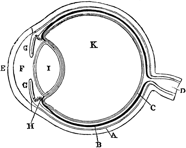
Diagram of the Eye | ClipArt ETC
Eye Anatomy: A Closer Look At the Parts of the Eye - All About Vision The iris of the eye functions like the diaphragm of a camera, controlling the amount of light reaching the back of the eye by automatically adjusting the size of the pupil (aperture). The eye's crystalline lens is located directly behind the pupil and further focuses light.
Labeled Picture Of The Eye - ClipArt Best
Human Heart - Anatomy, Functions and Facts about Heart - BYJUS The human heart functions throughout a person’s lifespan and is one of the most robust and hardest working muscles in the human body. Besides humans, most other animals also possess a heart that pumps blood throughout their bodies. Even invertebrates such as grasshoppers possess a heart like pumping organ, though they do not function the same way a human heart …
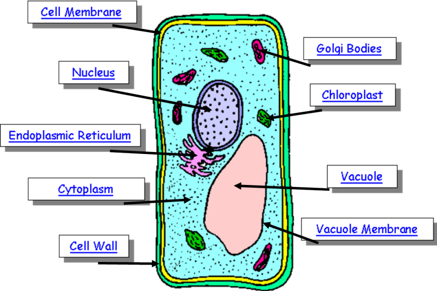
Plant Cell Drawing With Labels at PaintingValley.com | Explore collection of Plant Cell Drawing ...
plotly.com › python › parallel-categories-diagramParallel categories diagram in Python - Plotly Basic Parallel Categories Diagram with graph_objects¶ This example illustrates the hair color, eye color, and sex of a sample of 8 people. The dimension labels can be dragged horizontally to reorder the dimensions and the category rectangles can be dragged vertically to reorder the categories within a dimension.

Learning English in a new way: diciembre 2013
PDF Eye Anatomy Handout - National Institutes of Health of light entering the eye. Lens: The lens is a clear part of the eye behind the iris that helps to focus light, or an image, on the retina. Macula: The macula is the small, sensitive area of the retina that gives central vision. It is located in the center of the retina. Optic nerve: The optic nerve is the largest sensory nerve of the eye.

Main veins of the leg stock vector. Illustration of iliac - 121325793
Labeled Eye Diagram - Pinterest This vibrant 20" x 26" (51 x 66 cm) exam-room anatomy poster shows cross section of The Eye. It also provides lateral and superior view of the eye and shows the visual field. Anterior chamber angle, eyelashes, tear ducts, cornea, lens, retina, fundus and the macula lutea are illustrated.

Draw a labelled diagram of the human eye. Label the following parts on this diagram:
byjus.com › biology › liver-diagramLiver Diagram with Detailed Illustrations and Clear Labels Liver – Anatomy, Functions, And Liver Diseases; Also Read: 6 Facts Everyone Should Know About The Liver; Fatty Liver Symptoms – Explore The Signs, Indications And Causes; 12 Alarming Symptoms of Liver Problems You Shouldn’t Ignore; A Brief Account Of Hepatic Portal System And Its Significance; Human Body – Anatomy and Physiology of ...
Document Moved
Structure And Function Of The Eye - Vision - MCAT Content - Jack Westin Structure and Function of the Eye. The human eye is an organ that reacts with light and allows light perception, color vision, and depth perception. The photoreceptive cells of the eye, where transduction of light to nervous impulses occurs, are located in the retina (shown in Figure 1) on the inner surface of the back of the eye.

Human Eye Diagram Labeled - Health, Medicine and Anatomy Reference Pictures | School | Pinterest ...
Structure and Functions of Human Eye with labelled Diagram - BYJUS Structure and Functions of Human Eye with labelled Diagram Biology Biology Article Structure Of Eye Structure of the Eye The eye is one of the sensory organs of the body. In this article, we shall explore the anatomy of the eye The structure of the eye is an important topic to understand as it one of the important sensory organs in the human body.

Module 1: Labeled Diagram of the Eye | Eye health | Pinterest
The Eye Diagram: What is it and why is it used? The eye diagram is used primarily to look at digital signals for the purpose of recognizing the effects of distortion and finding its source. To demonstrate using a Tektronix MDO3104 oscilloscope, we connect the AFG output on the back panel to an analog input channel on the front panel and press AFG so a sine wave displays. Then we press Acquire.
The eye, rods and cones - Biology Notes for IGCSE 2014
Eye Anatomy Diagram - EnchantedLearning.com Retina - light-sensitive tissue that lines the back of the eye. It contains millions of photoreceptors (rods and cones) that convert light rays into electrical impulses that are relayed to the brain via the optic nerve. Rods - cells the in the retina that sense brightness (they are photoreceptors). Night vision involves mostly rods (not cones).

Functions and Anatomy of the Eye by Health EDventure | TpT
Label Parts of the Human Eye - University of Dayton Parts of the Eye. Select the correct label for each part of the eye. The image is taken from above the left eye. Click on the Score button to see how you did. Incorrect answers will be marked in red. ...
Plant cell Structure: Plant cell parts, Organelles and their functions and Diagram - Jotscroll
Cow's Eye Dissection - Eye diagram - Exploratorium The pupil is the dark circle in the center of your iris. It's a hole that lets light into the inner eye. Your pupil is round. A cow's pupil is oval. A tough, clear covering over the iris and the pupil that helps protect the eye. Light bends as it passes through the cornea. This is the first step in making an image on the retina.

Can anyone pls help me with an eye (fully labelled)diagram...it's given wrong in our book.Kindly ...
Label the microscope — Science Learning Hub 08.06.2018 · All microscopes share features in common. In this interactive, you can label the different parts of a microscope. Use this with the Microscope parts activity to help students identify and label the main parts of a microscope and then describe their functions.. Drag and drop the text labels onto the microscope diagram. If you want to redo an answer, click on the box and …
Structure And Function Of The Eye - YouTube
Parallel categories diagram in Python - Plotly Multi-Color Parallel Categories Diagram¶. The color of the ribbons can be specified with the line.color property. Similar to other trace types, this property may be set to an array of numbers, which are then mapped to colors according to the the colorscale specified in the line.colorscale property.. Here is an example of visualizing the survival rate of passengers in the titanic …
Easy Human Eye Diagram With Labels - Diagram Media
Structure of Human Eye (With Diagram) | Human Body - Biology Discussion It extends from the ciliary body across the eyeball in front of the lens. It has an opening in the centre called the pupil. It contains two types of smooth muscles, circular muscles (sphincters) and radial muscles (dilators), of ectodermal origin. The iris controls the amount of light entering the eye by the radial muscles contracting in dim ...

Diagram Of An Eye And Its Functions - Diagram Media
MCAT Eye Anatomy: Eye Structure & Function - Magoosh MCAT Blog Function. Cornea. Outermost lens of the eye responsible for the majority of light refraction. Aqueous humor. Fluid that adds refractive power. Iris/Pupil. Colored tissue that regulates the amount of light entering the eye through the central pupil. *KEY CONCEPT: The pupil dilates in low light conditions and constricts in high light conditions.
Post a Comment for "43 eye diagram with labels and functions"